


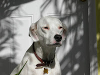
 Boo is a 5-year-old Dalmatian mix whose owners came in to have a soft mass on his right front leg evaluated. Eight months previously, they had brought Boo in for his annual exam at another area animal hospital. They had pointed out the mass to the veterinarian at that time and were told that it “felt like a benign fatty tumor” and not to worry about it. Since that first exam, the lump had doubled in size, so they came to Friendship to get a second opinion.
A fine needle aspirate is the only way to know for sure whether a suspicious lump is a benign fatty tumor (also called a lipoma), or a malignant growth. It is a simple, virtually painless procedure in which a needle is inserted into the mass so that a few cells can be obtained for examination under the microscope. Boo was very brave as I performed the aspirate. In fact, he didn’t seem to notice I was doing anything!
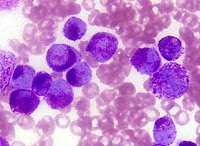 MCTs are the most common type of canine skin cancer, making up about 20% of all skin tumors. They have a wide variety of behavior. Some are easily cured by surgical excision, while others metastasize to lymph nodes, liver or spleen.
The first step for Boo was to schedule surgery to have the mass removed so we could determine his prognosis and treatment options. MCTs are graded by a pathologist on a scale of 1 to 3, with grade III being the most malignant.
Given the size and location of Boo’s mass, it was going to be challenge to remove all of it, along with the extra tissue required to get what we call “clean margins.” Lesions on an animal’s leg or face are much more difficult to remove as you are limited by the lack of excess skin to work with.
The fact that the tumor had grown so much bigger also decreased the likelihood that we’d be able to remove it completely. If it had been discovered to be an MCT during the first visit at the other animal hospital, surgery could have been performed early on while the tumor was still small and the chances were greater it could be removed completely. But because a fine needle aspiration was not performed, the tumor was not correctly diagnosed and was instead allowed to double in size.
Normally I would have no problem surgically removing an MCT, but given the location of Boo’s mass I referred him to our board-certified surgeon Dr. Walker. Though Dr. Walker removed as much tissue surrounding the mass as possible, he was not confident that he was able to get clean margins. To find this out, we would have to wait for the pathology report.
Boo’s MCT came back as a Grade II with dirty margins, meaning we had not been able to remove all of the tumor cells. The next step was to send Boo to the oncology department for more extensive diagnostics to determine if the cancer cells had spread anywhere else.
Boo was lucky. The MCT had not spread and he was an excellent candidate for a new drug called Palladia, the very first chemotherapy drug approved by the FDA for use in veterinary medicine. I am also proud to report that The Oncology Service at Friendship performed many of the central clinical trials that were use in gaining FDA approval of this medication. Normally veterinarians and veterinary oncologists use human drugs off-label so the release of Palladia is an exciting step in the fight against canine cancer.
The lesson I hope that everyone can learn from Boo’s story is that ALL skin masses should be aspirated, even if they “feel” benign. I am confident that Boo is in the very best hands with our oncology department and currently he is doing very well.


No comments:
Post a Comment